Keratoconus: Early Signs, Diagnosis and Monitoring
Frequent prescription changes or distorted vision? Learn how keratoconus is diagnosed, why monitoring matters and when cross-linking is considered.
Kiran Challa
10/22/20263 min read

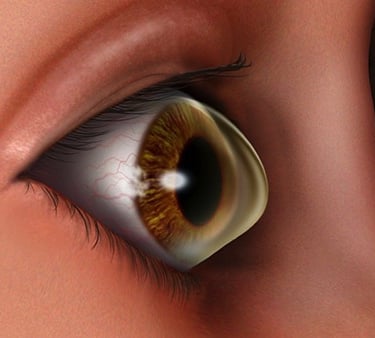
Frequent changes in glasses, ghost images or blurred vision that remains after a new prescription can be reasons to investigate the cornea. They do not automatically mean keratoconus, but they deserve an eye examination.
Keratoconus is a condition in which the cornea (the clear front surface of the eye) becomes thinner and develops an irregular, cone-like shape. It often starts in adolescence or early adulthood. Progression varies, so early diagnosis and repeat measurements are important.
Early signs to look out for
Repeated changes in spectacle prescription, especially astigmatism.
Blurred or distorted vision despite updated glasses.
Ghost images, glare, halos or difficulty seeing clearly at night.
Light sensitivity, squinting or visual strain.
A family history of keratoconus, frequent eye rubbing and persistent eye allergy are relevant risk factors to discuss. They are not symptoms or proof of the condition.
Why does the cornea change shape?
The cause is not fully understood. Genetic susceptibility and biological and environmental factors can contribute. Eye rubbing is an important modifiable risk factor associated with development or progression.
If your eyes itch, seek advice about managing allergy or irritation rather than rubbing them repeatedly. Keratoconus commonly affects both eyes, often to different degrees.
How is keratoconus diagnosed?
Refraction and vision assessment
The clinician measures how well glasses or lenses correct vision. An unusual or changing prescription may prompt further investigation, but refraction alone cannot establish the diagnosis.
Slit-lamp examination
This examination allows close inspection of the cornea for thinning, scarring and other changes. Early disease may require imaging even when obvious signs are absent.
Corneal topography and tomography
Topography maps corneal curvature. Tomography provides additional information about the front and back surfaces and the distribution of corneal thickness. These measurements help identify patterns suggestive of keratoconus.
Corneal thickness measurement
Pachymetry measures thickness and helps guide assessment and treatment planning. No single thickness value or map should be interpreted in isolation.
How do clinicians assess progression?
The clinician compares the overall findings with previous examinations, including changes in corneal shape, thickness, refraction and vision. A single scan is useful as a baseline, but repeated measurements help reveal whether the condition is changing.
Children and younger adults may require closer monitoring. The appropriate interval depends on age, clinical findings and suspected progression.
Bring previous scans and prescriptions where possible. Tell the clinic about contact lens wear before imaging; your clinician may ask you to leave lenses out for a specified period because lens wear can affect corneal measurements.
Vision correction and progression control
These are separate goals. Glasses or contact lenses help focus light, while corneal collagen cross-linking aims to strengthen the cornea and reduce further progression.
Early disease: glasses or suitable soft lenses may provide adequate clarity.
Irregular corneal optics: corneal rigid, hybrid or scleral lenses may improve functional vision.
Progressive disease: an ophthalmologist may recommend cross-linking after assessing suitability.
Advanced disease: significant scarring or inadequate vision may require surgical assessment, including consideration of ring segments or transplantation in selected cases.
Cross-linking is not a guaranteed way to restore normal vision or remove the need for lenses. Likewise, clear vision through a scleral lens does not mean progression has stopped.
For detailed fitting information, read Scleral lenses for keratoconus: vision, comfort and care.
When to seek prompt care
Arrange an examination for recurring visual changes or difficulty obtaining clear vision with glasses. Sudden marked blur, eye pain, significant redness or a cloudy-looking cornea needs prompt clinical assessment rather than waiting for a routine review.
Frequently asked questions
Can a normal glasses test rule out early keratoconus?
Not always. Suspicious symptoms or findings may justify corneal imaging and follow-up even when useful vision can still be achieved with glasses.
Does everyone with keratoconus need surgery?
No. Many people use glasses or specialty lenses for vision. Cross-linking or other procedures are considered according to progression, corneal health and visual needs.
Can keratoconus cause serious vision loss?
Advanced thinning and scarring can cause substantial visual impairment. Assessment and appropriate management help address progression and preserve functional vision.
Can I stop reviews once my vision feels stable?
Keep the agreed review schedule. Changes in the cornea may not be obvious from symptoms alone.
Arrange an assessment
Contact Nexus Vision Eye Care in Secunderabad to discuss an examination. Bring your current glasses, contact lens details and earlier prescriptions or corneal scans.
Further reading
Mayo Clinic: keratoconus diagnosis and treatment.
About the author
Kiran Challa, Specialist Optometrist. His clinical interests include specialty contact lenses for complex corneal conditions and orthokeratology for myopia management.
Updated 22 October 2026. This article is for patient education and does not replace an individual eye examination or professional advice.
Subscribe to our newsletter
Contacts
+91 8247710054
nexusvisioneyecare@gmail.com